Ovary
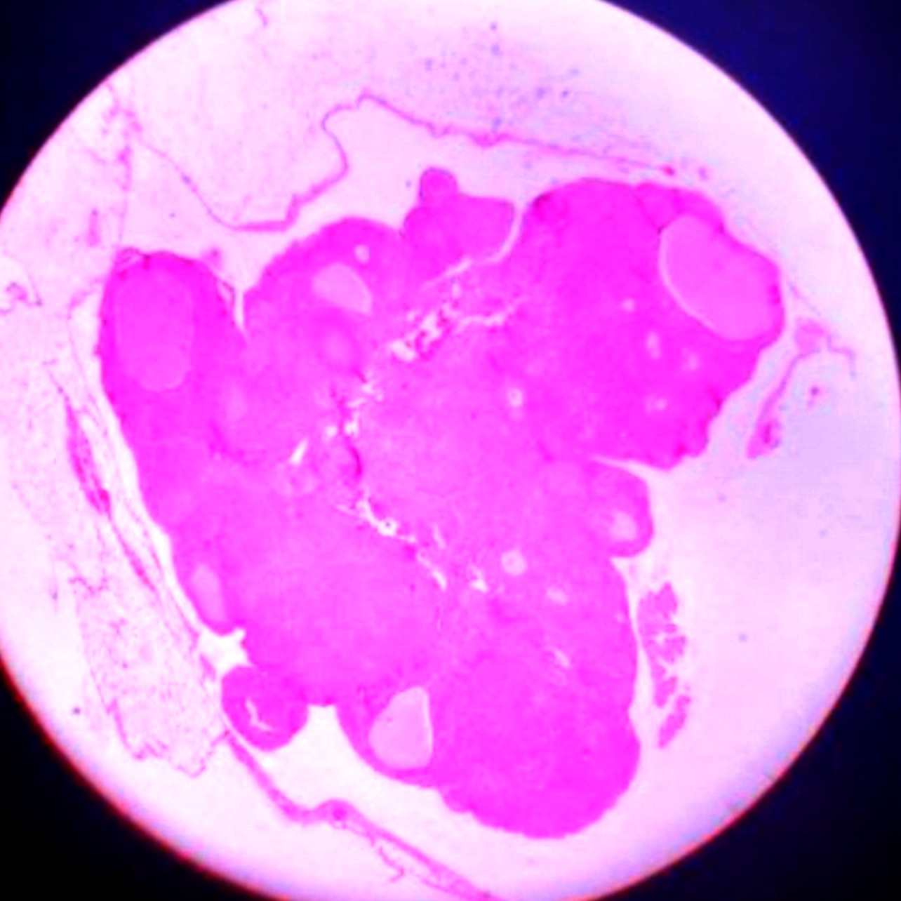
Identification Points
1. Outer cortex-shows ovarian follicles in different
stages of development,primordial, primary,
secondary and Graafian follicle.
2. Inner medulla-contains connective tissue
with blood vessels.
Fallopian Tube

Identification Points
1. Muscular tube.
2. Mucus layer-thrown into numerous
branching and anastomosing folds
which almost fill the lumen.
3.Lined by columnar cells of two types:
Ciliated - which help in movement of
ova towards the uterine cavity.
Non-ciliated - secretory , secretions help
in nourishment of ova.
Umbilical Cord

Identification Points
1. Two muscular arteries and one vein seen.
2. Wharton's jelly (embryonic mesenchyme)
surrounding the vessels with outer
amniotic covering.
Placenta

Identification Points
1. Sections of chorionic villi seen.
2. Villi with embryonic connective tissue core
containing fetal capillaries and surrounded
by outer syncytiotrophoblasts and
inner cytotrophoblasts.
Uterus - Secretory

Identification Points
1. Inner endometrium-lined by simple
columnar cells.
Stroma contains tubular uterine glands
and spiral arteries.
2. Middle thick muscular layer-myometrium.
3. Outer perimetrium.
Uterus - Proliferative

Identification Points
1. Inner endometrium-lined by simple
columnar cells.
Stroma contains tubular uterine glands
and spiral arteries.
2. Middle thick muscular layer-myometrium.
3. Outer perimetrium.